

Female ribcage Stock Image P116/0678 Science Photo Library
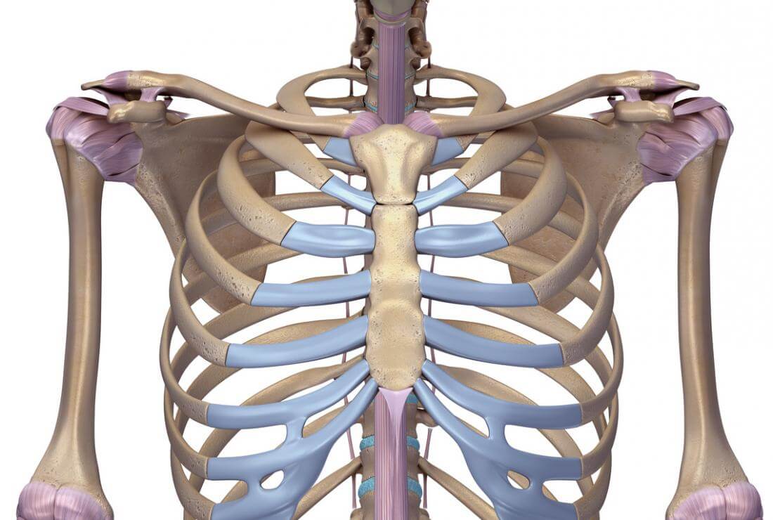
The rib cage surrounds the lungs and the heart, serving as an important means of bony protection for these vital organs.In total, the rib cage consists of the 12 thoracic vertebrae and the 24 ribs, in addition to the sternum. With each succeeding rib, from the first, or uppermost, the curvature of the rib cage becomes more open.
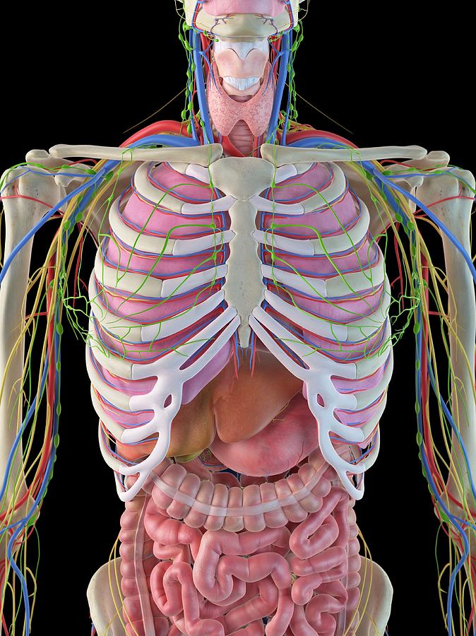
Diagram Rib Cage With Organs / Chest bone rib cage landmark diagram
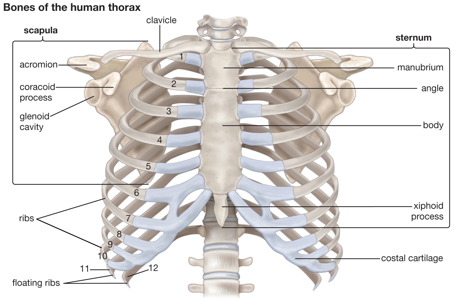
The ribs are curved, flat bones which form the majority of the thoracic cage.They are extremely light, but highly resilient; contributing to their role in protecting the internal thoracic organs. There are twelve pairs of ribs, all of which articulate with the vertebral column.However, only seven have a direct articulation with the sternum.As such, ribs can be allocated to one of three.

Female Rib Cage Image & Photo (Free Trial) Bigstock
Skeleton & Spine Shoulder & Back Arm & Hand Pelvis & Hip Leg & Foot has three important functions: protection, support and respiration. The bones of the rib cage are the thoracic vertebrae and the 12 pairs of is a flat bone that is made up of three parts, the (1) manubrium,, and the ( 3) xiphoid process.

Rib Cage Medical Art Library
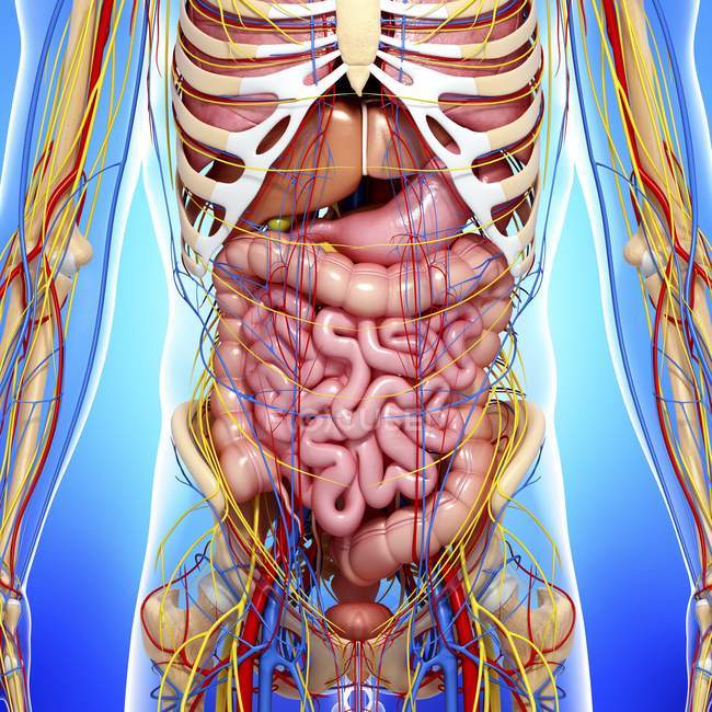
In human body, the rib cage is a basket-like structure that is formed from the ribs and their corresponding attachments to the sternum and vertebral column. The rib cage structure houses two vital organs, the lungs and the heart and provides them with bony protection from outside injury and trauma. The rib cage is expansible and semi-rigid in.
Rib Cage Yoga and Medical Science
Adding text and lines: Derive "from scratch"[edit] By this method, body diagrams can be derived by pasting organs into one of the "plain" body images shown below. This method requires a graphics editor that can handle transparent images, in order to avoid white squares around the organs when pasting onto the body image.

Female body anatomy, lung organ, gastrointestinal organs png PNGEgg
These internal structures of female anatomy include the: Vagina: The vagina is a muscular canal that connects the cervix and the uterus. It leads to the outside of the body. Parts of the vagina are made of collagen and elastin, which help it expand during sexual stimulation and childbirth. Cervix: The cervix is the lower part of the uterus that.

Xray View Of Female Chest Rib Cage Heart Arteries Veins Anatomy Stock
The thoracic cage, also known as the rib cage, is the osteocartilaginous structure that encloses the thorax.It is formed by the 12 thoracic vertebrae, 12 pairs of ribs and associated costal cartilages and the sternum.. The thoracic cage takes the form of a domed bird cage with the horizontal bars formed by ribs and costal cartilages. It is supported by the vertical sternum (anteriorly) and the.
Picture Of What Is Under Your Rib Cage The spleen sits under your rib
The Ribs: Location, Anatomy, Functions, & Labeled Diagram Ribs Home Chest Bones Ribs Published on November 18th 2022 by staff What Are Rib Bones The ribs are 12 pairs of curved, flat bones that form the thoracic cage or rib cage, the bony structure that shapes the thoracic cavity and protects various organs.

de Female Human Anatomy Organs Diagram mar webmds abdomen anatomy page
Edited By: Sagar Aryal. The rib cage or thoracic cage (called thorax) is located in the most medial portion of the body and protects vital organs like the heart and lungs. The rib cage is an integral part of the axial skeleton in vertebrates and it lies in the thoracic cavity. Some organs of the respiratory, cardiovascular, nervous, immune, and.

Human Rib Cage, Human Skeleton Anatomy, Human Body Anatomy, Human
The thoracic cage, a flexible framework of bones and cartilage, is conical in shape. It is narrower at the top and broadens to fit and protect some critical organs of respiration and circulation—that is, the lungs and heart. The thoracic cage gives your upper torso structure. Women have smaller cages than men; the capacity is less, and the.

Female rib cage and spine. Stocktrek Images
Articulations Xiphisternal joint - Between the sternum's body and xiphoid process Manubriosternal joints - Between the sternum's body and the manubrium Sternoclavicular joints - Between the manubrium (top of the sternum) and the clavicles Sternochondral joints - Between the sternum and the costal cartilage
Anatomy Rib Cage Organs / Lungs And Rib Cage Stock Illustration
From Wikipedia, the free encyclopedia , which protects . The circumferential enclosure formed by left and right rib cages, together known as the thoracic cage, is a semi-rigid structure which surrounds the thoracic cavity and supports the shoulder girdles axial skeleton

Image result for Rib Human ribs, Body anatomy, Anatomy bones
Ribs 1, 2, 10 11 and 12 can be described as 'atypical' - they have features that are not common to all the ribs. Rib 1 is shorter and wider than the other ribs. It only has one facet on its head for articulation with its corresponding vertebra (there isn't a thoracic vertebra above it).

Anatomy Diagram Rib Area / Anatomy Of The Female Abdomen And Pelvis
Summary. The five vital organs in the human body are the brain, heart, lungs, kidneys, and liver. Other organs include the gallbladder, pancreas, and stomach. Organ systems, such as the nervous.
What are the functions of rib cage? Information About Rib Cage
Rib cage | BioDigital Anatomy Skeletal system > Axial skeleton > Rib cage This image was produced from interactive 3D models on the BioDigital Human Interact in 3D Rib cage Interactive 3D Anatomy The BioDigital Human - Platform Overview

Illustration. Human rib cage over lungs, heart, other lower intestine
Figure 7.5.1 - Thoracic Cage: The thoracic cage is formed by the (a) sternum and (b) 12 pairs of ribs with their costal cartilages. The ribs are anchored posteriorly to the 12 thoracic vertebrae. The sternum consists of the manubrium, body, and xiphoid process. The ribs are classified as true ribs (1-7) and false ribs (8-12).